Bella is a sweet and gentle 10 year old female Australian Shepherd mix that was hiding a problem so severe that it would cause any human with a similar disease process to be expressing absolute misery. Yet, Bella was outwardly a happy and active dog who was well taken care of and very loved. Bella illustrates how well our beloved pets can hide even severe oral pain and how appropriate treatment can stop the pain and let them truly enjoy life.
Bella and her family recently moved to Asheville from Tennessee. Her mom, Holly, was concerned because in spite of having professional dental cleanings every six months and having excellent oral homecare including daily tooth brushing, Bella developed severe halitosis and bleeding from her gums within two months after professional cleaning.
At her initial visit here at Animal Hospital of North Asheville, Dr. Earley called me in to evaluate the inflammation seen in Bella’s mouth.

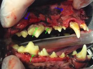
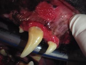
Bella had severe inflammation of her oral cavity consistent with a disease process called Chronic Ulcerative Paradental Stomatitis (CUPS) as well as severe, end stage periodontal disease. See pictures 1 and 2.


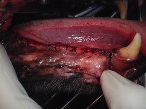
Unlike periodontal disease which affects and destroys the gingiva (gums), the periodontal ligament (lining the tooth socket), the alveolar bone (forms the socket) and the cementum (covering of the tooth root), CUPS affects any tissue in the mouth that comes in contact with plaque or tartar (including the inside of the mouth and tongue). Sores called “kissing lesions” (See picture 3) are common.

The disease is characterized by SEVERE inflammation of tissues and occurs due to the pet’s immune system becoming hypersensitive to plaque. CUPS is reported to occur more frequently in certain breeds of dogs like Maltese, Cavalier King Charles, and Greyhounds, but Dr. Thompson and I have seen the disease in many other breeds of dogs including mixed breeds.
CUPS can develop slowly over years or suddenly, and as was the case with Bella, often concurrently causes severe periodontal disease. A dog with CUPS may exhibit severe halitosis, drooling, bleeding from the mouth and gums, reluctance to have their mouth or muzzle touched, reluctance to play with toys, difficulty eating, dropping of food, or decreased food intake.
The disease is diagnosed by having a thorough oral examination, ruling out other disease processes, and the possible biopsy of affected tissue. In mild or early cases, aggressive prevention of plaque buildup on the teeth may be an effective treatment.
However, this can prove quite difficult as it requires semi-annual professional cleanings, a minimum of once daily tooth brushing (twice daily is more effective), the application of a tooth sealant called Oravet, the use of dental diet and treats, and the use of an oral antibacterial rinse. Unfortunately, despite attempts at preventing plaque buildup, CUPS often persists or progresses. In these cases, the best treatment is extraction of some or often all the teeth in the mouth that provide a surface for plaque to accumulate on.
While this may sound extreme, this disease is extreme, and the quality of life of a pet with CUPS can be dramatically improved with this appropriate treatment. Unfortunately, Bella’s veterinarian(s) in Tennessee did not diagnose the disease, and while cleanings were performed, the disease continued to progress. The necessary treatments were not discussed with Holly and Bella continued to suffer.
After Bella’s initial evaluation here, a more thorough oral evaluation under anesthesia was scheduled. Very typically, these patient’s mouths are so painful that the mouth cannot be examined at all unless they are under anesthesia.
I found Bella to have severe inflammation of her oral tissues AND end stage periodontal disease. (See pictures 1 and 2.) In Bella’s case, it was obvious that good homecare and professional cleanings were not enough to control her disease and the decision was made that the best treatment for Bella was to extract all her teeth. Due to the extensive nature of the surgery, it was done in two separate procedures.


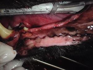
The resulting rewards are obvious. Pictures 4 and 5 (above) demonstrate how Bella’s mouth looked immediately after the extractions were performed and pictures 6 and 7 (below) show her mouth once it was healed. What a dramatic difference!! Also note that the large fang that remained after her first surgery, seen in picture 6, was still inciting a large degree of inflammation. This tooth was extracted during her second surgical visit.


After the first surgery was performed, Holly stated that Bella had a very good recovery and appeared to have more energy than previously. After the second stage of the surgery was performed, Holly stated that Bella was "bounding around like a two year old!” While Holly understandably had concerns about the procedure beforehand, she is very happy with the outcome and Bella is happier and healthier than ever.
Aggressive pain control is used post-operatively and dogs undergoing such a procedure have a comfortable, rapid recovery. Once the extraction sites are healed, many dogs with no or few teeth are able to still eat dry kibble, play with toys, and lead very normal (and better) lives.
We are all very happy for Bella and her family!! Please allow us to evaluate your dog’s mouth at least once yearly so that we can ensure they have the best quality of life possible.
